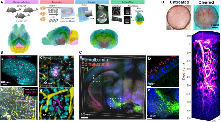
FIGURE 5.

Applications of tissue clearing in neuroscience. (A) Overview of construction of mouse brain atlas with single‐cell annotation (CUBIC‐Atlas), and the volume‐rendered images of the CUBIC‐Atlas from the horizontal, sagittal, and coronal view. (B) 3D visualization of amyloid plaques in AD using iDISCO clearing. a, Amyloid plaques and neurofilament H imaging in a cleared cortex slice of 10‐month‐old 2xTg AD mouse (500 μm thick). b, Maximum projection of vasculature, amyloid plaques, and microglia staining from an 11‐month‐old 2xTg AD mouse brain (1 mm thick). The insert is the magnification of the indicated region (white rectangle). c, The magnified optical section from the AD brain in b, the circles indicate the plaques surrounded by reactive microglia and vessels. d, The magnified surface render in c. (C) Multiplexed detection of mRNAs in EDC‐CLARITY. a, Multiplexed in situ hybridization of coronal section (0.5 mm thick) using somatostatin (red), parvalbumin (blue), and tyrosine hydroxylase (green) probe sets. b, c, Magnified view of indicated boxes. (D) 3D imaging of mouse brain vasculature labeled with Texas Red through the transparent skull window. Images adapted from Refs (Liebmann et al., 2016; Sylwestrak et al., 2016; Murakami et al., 2018; Chen et al., 2019)
