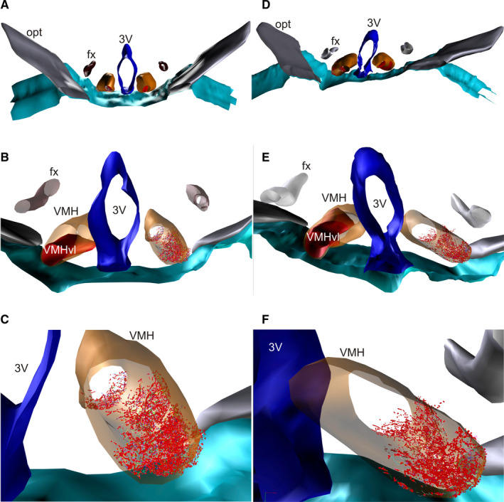
FIGURE 5.

Schematic 3D reconstruction of PHA‐L‐labelled terminals in the VMHvl used for the quantification of fibre and varicosity numbers. Labelled terminals in the VMHvl (in red) after PHA‐L injection in the MPN of control (a–c) and OVX rats (d–f). 3V, third ventricle; fx, fornix; opt, optic tract; VMH in orange
