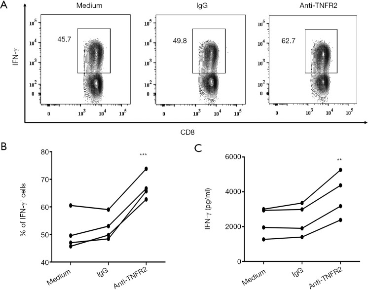
Figure 6.
Effects of blocking TNFR2 on effector CD8+T cell function present in MPE. PEMCs from MPE were cultured in medium alone, anti-TNFR2 mAbs, or isotype IgG. On day 3, intracellular staining was performed following a 5-hour stimulation of PEMCs with PMA (50 ng/mL) and ionomycin (1 µg/mL). Four hours into stimulation, Brefeldin A was added at a final concentration of 3 µg/mL to block cytokine secretion. (A) Representative FACS analysis of IFN-γ+ cells in CD8+T cells after 72 h at the indicated conditions, and (B) summary data are shown (n=4). (C) Comparison of IFN-γ concentrations in the 72-h supernatants of PEMCs exposed to anti-TNFR2 mAbs or Isotype IgG (n=4). Flow analysis was gated on live CD3+CD8+ cells. Data are expressed as means ± SEM; **, P<0.01, ***, P<0.001 by paired Students t-test. TNFR2, tumor necrosis factor receptor type II; MPE, malignant pleural effusion; PEMC, pleural effusion mononuclear cell.

