Figure 1.
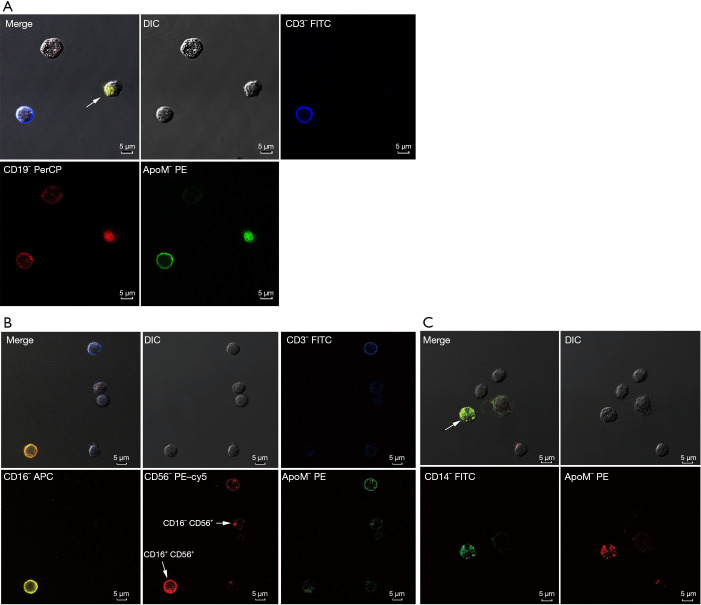
Confocal fluorescence microscopic images of apoM+ cells in PBMCs. Localization was carried out by incubation of cells with rabbit anti-human apoM or rabbit IgG (isotype control). (A) ApoM is expressed on CD19+ B cells (marked with an arrow). Blue: CD3+; red: CD19+; green: apoM+. (B) ApoM is expressed on CD3+ T cells and the CD16+ CD56+, the CD16– D56+ NK cells (marked with arrows). Blue: CD3+; yellow: CD16+; red: CD56+, green: apoM+. (C) ApoM is expressed on CD14+ monocyte (marked with an arrow). Green: CD14+; red: apoM+. ApoM, apolipoprotein M; PBMCs, peripheral blood mononuclear cells. Merge, a overlapped image from multiple fluorescence channels. DIC, differential-interference-contrast image.

