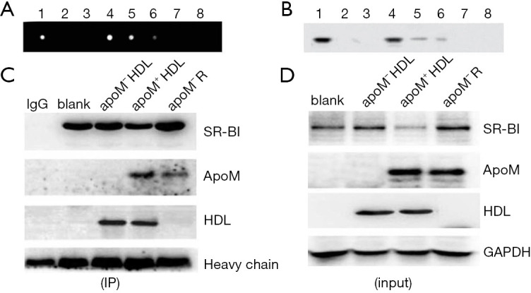
Figure 3.

Identification of the interactions between SR-BI and apoM by co-immunoprecipitation. (A,B) Dot blot and Western blot analysis of apoM in effluent liquid and elution fractions. 1: unpurified HDL (diluted with PBS 1:10); 2: effluent liquid (the flow-through after apoM+ HDL binding to the column, diluted with PBS 1:10); 3: wash liquid (the flow-through after washing with the wash buffer); 4–8: elution fractions (collected 0.5 mL/tube, 5 tubes). (C,D) SR-BI coimmunoprecipitated with apoM+ HDL, apoM− HDL, and recombinant apoM protein in THP-1 cells. THP-1 cells were incubated with 160 nM PMA and 100 µg/mL ox-LDL for 24 hours, respectively. Fifty µg/mL of both apoM+ HDL and apoM− HDL, as well as 1 µg/mL of recombinant apoM protein, was added to the medium for 6 hours before cell harvest. Cell lysates were immunoprecipitated using SR-BI antibodies coated with protein A beads. Immunoprecipitates were resolved by denaturing SDS-PAGE, then blotted and probed with HDL and apoM antibodies. apoM, apolipoprotein M; SR-BI, scavenger receptor BI; HDL, high-density lipoprotein; PBS, phosphate buffer saline.
