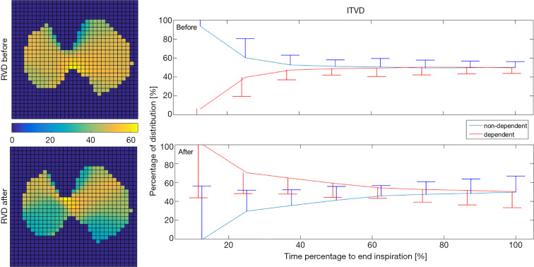
Figure 2.
Regional ventilation delay (RVD; left) and intra-tidal ventilation distribution (ITVD; right) analysis of the same patient as in Figure 1. RVD maps reveal that the inspiration started soonest in the dorsal regions after bronchodilation (green regions in the left bottom image). ITVD analysis shows that the dorsal regions (gravity-dependent regions) fill faster during inspiration after bronchodilation.

