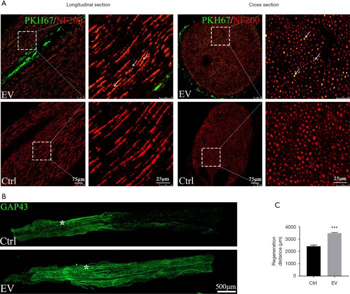
Figure 6.
Pro-regenerative effect of SKP-SC-EVs on a crushed nerve. (A) PKH67-labeled EVs (green) were locally injected into intact sciatic nerves. SKP-SC-EVs were co-localized with neurofilament (NF200, red) in the longitudinal and cross sections of the nerve tissue. Scale bar, 75 µm. Right panels correspond to higher magnification of the boxed areas. Scale bar, 25 µm. (B) Axonal regeneration of sciatic nerve with injection of EVs or PBS (control) in vivo. GAP43 (green) stained regenerating axons (from left to right) showed longer extensions in EVs group than that in control group. The proximal stump of the crush site was indicated by the asterisk. Scale bar, 500 µm. (C) Statistical histograms showed the average distances distal from the lesion site of the GAP43-positive nerve fibers 4 d after crush injury. Mean ± SEM, n=3, ***, P<0.001, as compared with control group. SKP, skin precursor; SC, Schwann cell; EV, extracellular vesicle.

