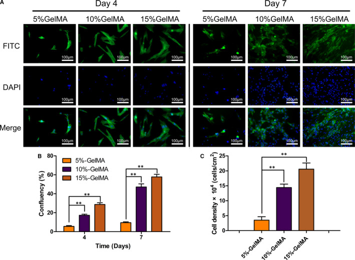
FIGURE 2.

Adhesion, proliferation and migration of NPCs on the GelMA hydrogel surface. (A) FITC‐labelled phalloidin /DAPI was used to stain actin filaments and nuclei of NPCs cultured for 4 and 7 days. (B) Over time NPCs’ confluence on GelMA hydrogel with different concentrations showed significant differences. (C) Cell density on the hydrogel surface on day 7 was measured and calculated by the number of DAPI staining nuclei positive in the restricted area. (*P < .05, **P < .01)
