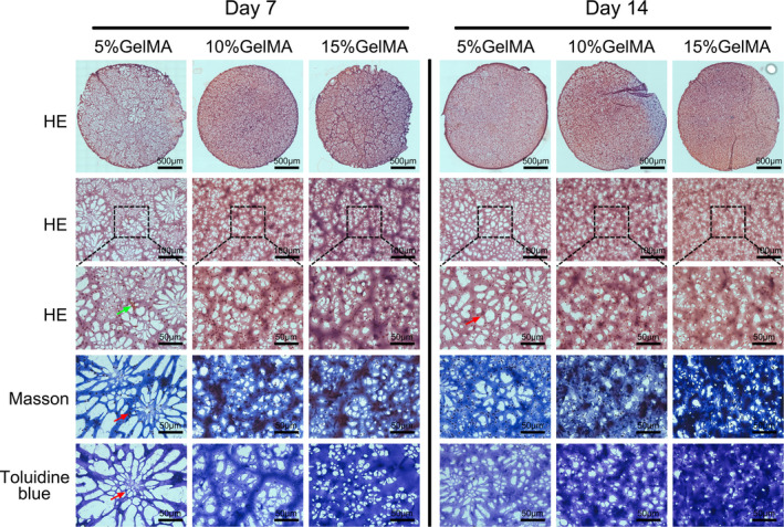
FIGURE 6.

Histological observation of GelMA hydrogel‐encapsulated NPCs. Micrographs showed H&E, Masson and toluidine blue staining of NPCs encapsulated in 5%, 10% and 15% (w/v) GelMA hydrogel on days 7 and 14 (green arrows indicating cell clusters; the red arrow indicates the single cell)
