Figure 1.

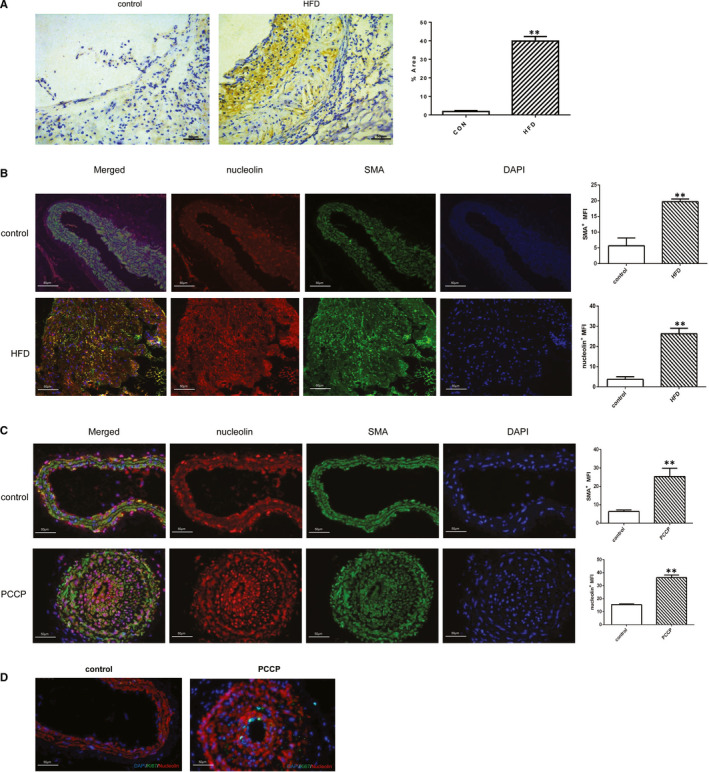
Nucleolin is up‐regulated during atherosclerosis development. (A) Representative immunostaining and quantification for nucleolin on aortic cross sections of ApoE–/– mice fed with chow and high‐fat diets. (Colorimetric images showing nucleolin in brown). Magnification 200×. HFD: high‐fat diet. **, P < .01, vs. control group, n = 3. (B) Immunofluorescence analysis showed co‐localization of nucleolin with SMA+ cells in ApoE–/– mice fed with chow and high‐fat diets. Immunostaining for nucleolin(red), SMA (green) and DAPI (blue). (C) Immunofluorescence analysis showed co‐localization of nucleolin with SMA+ cells in perivascular carotid collarp placement (PCCP) group and conrol group (n = 3). Immunostaining for nucleolin(red), SMA (green) and DAPI (blue). (D) Immunofluorescence analysis showed Ki67 with SMA+ cells in perivascular carotid collarp placement (PCCP) group and conrol group (n = 3). Immunostaining for SMA (red), Ki67 (green) and DAPI (blue)
