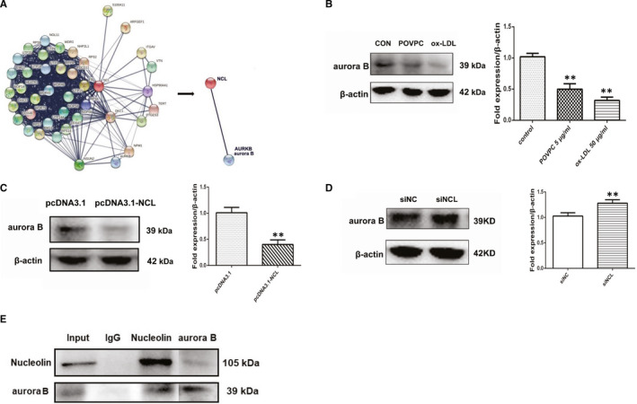
Figure 6.

Aurora B is a direct target of nucleolin in VSMCs. (A) Aurora B as a potential target gene by previous studies and String software (https://string‐db.org/). (B) The protein expression of aurora B fusion protein in vascular smooth muscle cells treated with POVPC or ox‐LDL. n = 3; **, P < .01 vs control group. (C) VSMCs were transfected with pcDNA3.1 or pcDNA3.1‐NCL. The protein level of aurora B was measured by Western blot. n = 3; **, P < .01 vs pcDNA3.1 (vector control). (D) VSMCs were transfected with siNCL or siNC. The protein level of aurora B was measured by Western blot. n = 3; **, P < .01 vs siNC (negative control). (E) Interaction of nucleolin and aurora B tested by immunoprecipitation. Lane 1 represented whole cell lysate. Lane2‐4 represented the proteins precipitated by control IgG, anti‐nucleolin, anti‐aurora B. The upper band indicated nucleolin, and the lower band indicated aurora B. Each experiment was repeated three times. P‐values were determined using the two‐tailed Student's t test for comparing two groups and one‐way ANOVA for comparing multiple groups
