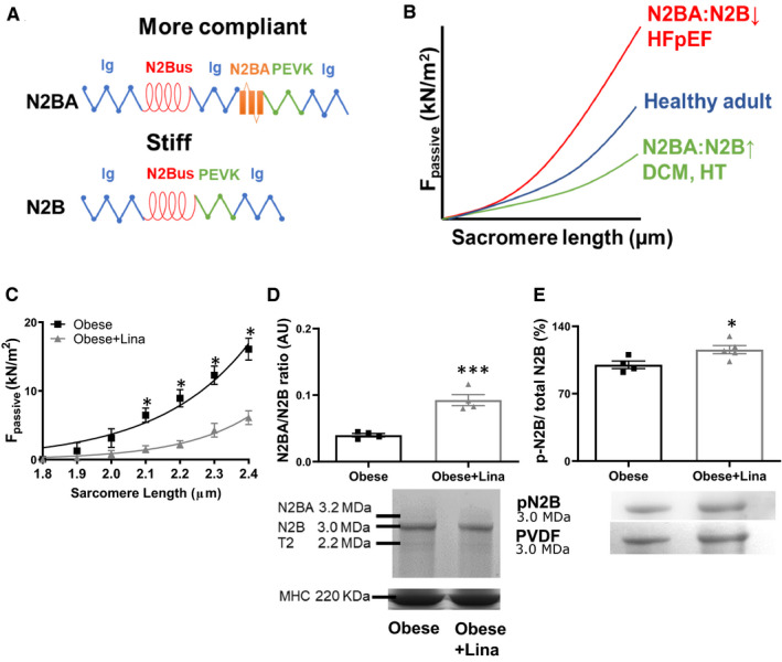
FIGURE 3.

Linagliptin induces isoform switching and total titin phosphorylation in obese ZSF1 rats. A, Structure composition of the extensible I‐band of the more compliant N2BA and stiffer N2B titin isoform. Blue, Ig domain; red, N2Bus domain; green, PEVK domain; orange, N2BA domain. B, Consequence of isotype switching on cardiomyocyte F passive. Titin isoform switching from the N2BA to the N2B isoform (reduced N2BA:N2B ratio) increases F passive, as observed HFpEF, while the inverse reduces F passive, as shown in dilated DCM and HT. C, Cardiomyocyte passive stiffness in isolated cardiomyocytes of linagliptin‐ (Obese + Lina) and placebo‐treated obese (Obese) ZSF1 rats at 20 wk (n = 3 different left ventricular tissues measuring at least 12 cardiomyocytes from each left ventricular tissue per condition). N2BA/N2B titin isoform ratio and representative Coomassie Blue stained‐PVDF membranes containing bands of N2BA, N2B and titin‐2 (T2; known degradation product), and loading control MHC (D) and total N2B titin phosphorylation and representative Coomassie Blue stained‐PVDF membranes (E) in linagliptin‐ (Obese + Lina) and placebo‐treated obese (Obese) ZSF1 rats at 20 wk (n = 4‐7 per group). DCM, dilated cardiomyopathy; F passive, passive stiffness; HFpEF, heart failure with preserved ejection fraction; HT, hypothyroidism; Ig, immunoglobulin; MHC, myosin heavy chain; N2Bus, N2B unique sequence. Data are expressed as mean ± SEM. All data were analysed using a two‐tailed unpaired Student t test with *P < .05 and ***P < .001
