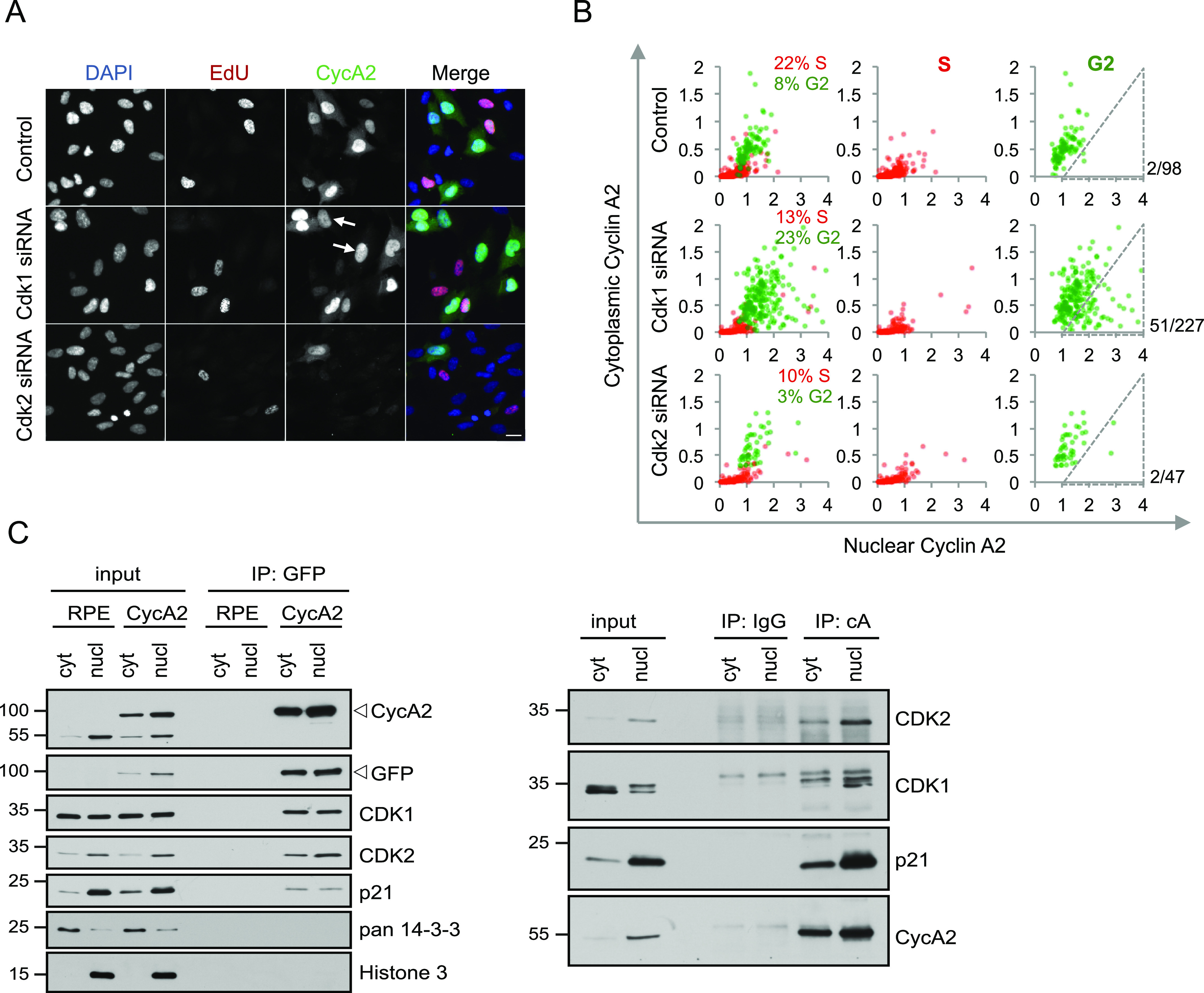
Figure 4. CDK1 can contribute to cytoplasmic accumulation of Cyclin A2.

(A) RPE cells were transfected with siRNAs for either CDK1 or CDK2 for 48 h, incubated with EdU for 20 min and fixed. Arrows indicate G2 cells with low cytoplasmic CycA2. Scale bar 20 μm. (B) Quantification of cytoplasmic and nuclear integrated intensities of CycA2 in at least 500 RPE cells imaged as in (A). Cells were gated for DAPI and EdU levels and assigned to the S phase (red dots) or G2 phase (green dots). Each dot represents one cell; the percentages indicate the proportion of the S- and G2 phase cells in each condition. Number of cells within indicated gate and total amount of G2 cells are shown to the right. (C) RPE (RPE) or RPE CycA2-eYFP (CycA2) cells were synchronised in G2, separated to cytosolic and nuclear fractions and immunoprecipitated with GFP Trap (left) or with control IgG and CycA2 antibody (right). Proteins bound to the carrier were probed with indicated antibodies. For (C), data using additional antibodies can be found in Fig 3E. All experiments were repeated at least three times.
