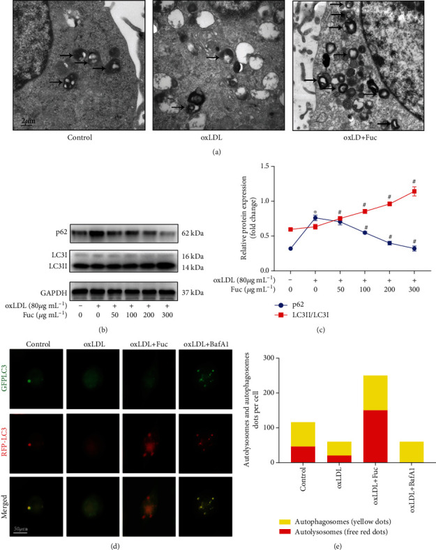
Figure 5.

Fucoidan enhances the impaired autophagy flux induced by oxLDL. (a) Autophagosomes in THP-1 cells detected by TEM. Autophagosomes are indicated by arrows. Scale bars = 2 μm. (b, c) Representative western blot analysis of the protein expression levels of LC3II/LC3I and p62. All data are representative of three independent experiments. (d) Fluorescence microscopy images of GFP-RFP-LC3 adenovirus transfected THP-1 cells treated with oxLDL, oxLDL+ fucoidan, or 50 nM baf A1 for 36 h. Scale bars = 50 μm. (e) The number of autolysosomes (red puncta) per cell was counted. Data are shown as the mean ± SE. ∗p < 0.05 vs. the untreated group; #p < 0.05 vs. the oxLDL-treated group.
