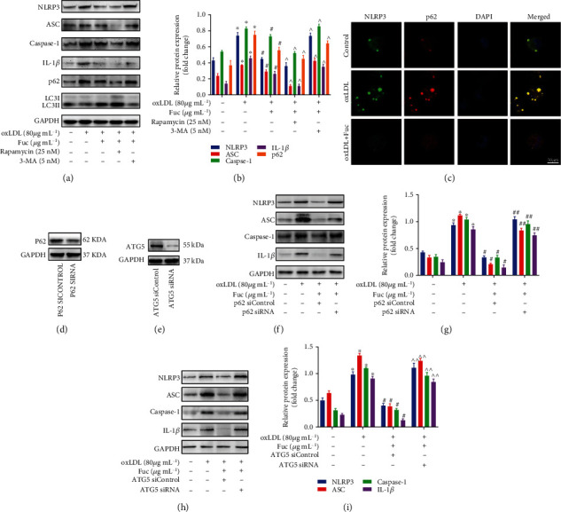
Figure 6.

Fucoidan mediates p62-dependent selective autophagy of NLRP3 inflammasome. (a, b) Representative western blot analysis of the protein expression levels of NLRP3 inflammasome-associated proteins, LC3II/LC3I, and p62. All data are representative of three independent experiments. (c) Fluorescence microscopy revealing the accumulation of NLRP3 and p62. Green indicates NLRP3 staining, red indicates p62 staining, and yellow indicates colocalization of NLRP3 and p62. Scale bar = 50 μm. (d) P62 protein expression following transfection with the control or p62 siRNA for 72 h. (e) ATG5 protein expression following transfection with the control or ATG5 siRNA for 72 h. (f–i) Representative western blot analysis of the protein expression levels of NLRP3, ASC, caspase-1, and IL-1β. All data are representative of three independent experiments. Data are shown as the mean ± SE. ∗p < 0.05 vs. the untreated group; #p < 0.05 vs. the oxLDL-treated group; ^p < 0.05 vs. the oxLDL + Fuc-treated group; ##p < 0.05 vs. the oxLDL+ Fuc + p62 siControl-treated group. ^^p < 0.05 vs. the oxLDL+ Fuc + ATG5 siControl-treated group.
