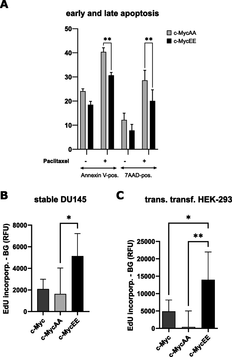
Fig. 6.

Effect of c-Myc phosphorylation mutants on apoptosis and proliferation. a Expression constructs of c-Myc with serines 67/71 mutated to alanines (preventing phosphorylation of these residues: c-MycAA), or mutated to glutamates (c-MycEE, mimicking IKKα-mediated phosphorylation) were stably transfected into DU145 cells. These cells were either left untreated or treated with 10 nM paclitaxel to stimulate apoptosis, followed by labeling with Annexin V (for early apoptotic cells) or 7AAD (for late apoptosis and necrosis) and measured by flow cytometry (n = 3, mean % positive cells +/−SD). b Proliferation of stable DU145 transfectants expressing c-Myc, c-MycAA or c-MycEE was assessed by culturing of cells in presence of 6 μM EdU for 4 h (to label cells in S-phase of the cell cycle). EdU was labeled by click-chemistry with TAMRA and the fluorescence was quantified on a plate reader with excitation at 546 nm and emission at 580 nm (n = 5, mean +/− standard deviation). c HEK-293 cells were transiently transfected with the c-Myc variants, followed by EdU-labeling of proliferating cells and quantification as described in (b). n = 6, Mean values are plotted with error bars indicating standard deviation
