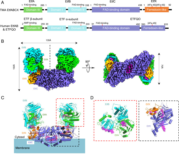
Fig. 1.
Superdimeric architecture of the Tma EtfABCX. (A) The domain architecture of EftA, B, C, and X in comparison with that of human EtfAB-QO. (B) Surface-rendered side and bottom views of the cryo-EM 3D map of the EtfABCX complex reconstituted by imposing a twofold symmetry. Subunits are individually colored with EtfA in green, EtfB in cyan, EtfC in purple, and EtfX in orange. (C) Cartoon views of the atomic model of EftABCX. The cofactors (three FAD molecules and two [4Fe-4S] clusters) are shown as magenta sticks superimposed with transparent spheres. Membrane is depicted as a cyan shade with the lipid-exposed and membrane-interacting hydrophobic side chains shown as sticks. (D) Zoomed views of EtfAB (Left) and EtfCX (Right); both are 90° rotated to better display the bound cofactors.

