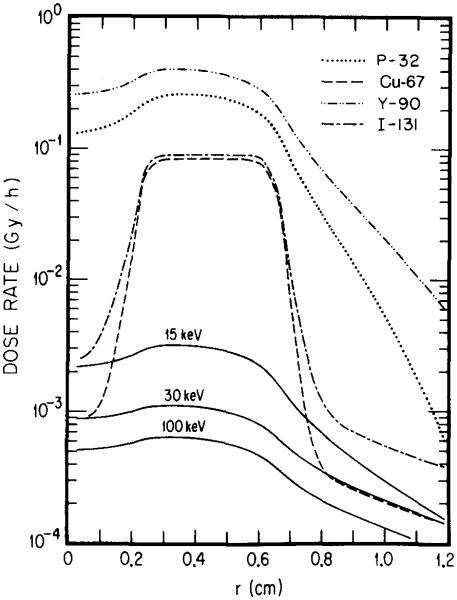
Fig. 8.
Spatial distribution of dose rates in the tumor model of Ref. 7 with a necrotic core. The tumor is a sphere of soft tissue, 0.66 cm in radius. The central region (0–0.22 cm) is necrotic with no uptake of radioactivity. Each one of the radionuclides, 32P, 67Cu, 90Y, and 131I, or a hypothetical monoenergetic photon emitter (either 15, 30, or 100 keV) is assumed to be uniformly distributed in the non-necrotic region of the tumor with a concentration of 1 MBq/ml. The region to the right of r = 0.66 cm is the normal tissue.

