Fig. 4.
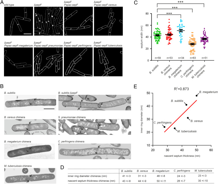
SepF ring diameter regulates septum thickness. (A) SIM images of B. subtilis ΔsepF cells expressing different SepF chimeras. The chimeras were expressed from the IPTG-inducible Pspac promotor. As a control we included the complementation strain expressing WT sepF from the same promoter. Cells were grown in the presence of 100 µM IPTG until midlog phase prior to membrane staining and microscopy. Lack of SepF results in severely deformed septa, which shows as highly fluorescent membrane patches due to membrane invaginations and double membranes. (B) Representative TEM images of septa of ΔsepF strains expressing the different SepF chimeras. (C) Quantification of the septum thickness of the different strains. Black bars represent mean with SEM. P values for B. megaterium, C. perfringens, and M. tuberculosis chimeras compared to B. subtilis are <0.0001. Number of measured septa is indicated in the graphs. Comparison (D) and correlation (E) of the inner ring diameter of SepF chimeras and nascent septum thickness of B. subtilis ΔsepF expressing the chimeras, or an IPTG-inducible WT sepF as control. Coefficient of determination is indicated above the graph. (Scale bars: A, 2 µm; B, 1 µm.)

