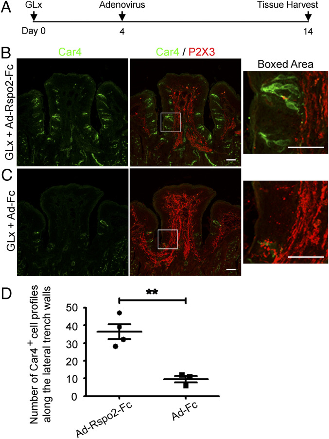
Fig. 4.
Generation of differentiated taste cells by Ad-Rspo2-Fc in the GLx mouse model. (A) Schematic of the experiment design. Adenoviral infection was performed on day 4 after GLx. (B) Immunostaining of sections from GLx mice infected with Ad-Rspo2-Fc shows numerous Car4+ cells (green) but no P2X3+ nerve terminal web (red) in taste buds in the circumvallate papilla. (C) Immunostaining of sections from GLx mice infected with control adenovirus (Ad-Fc) shows few Car4+ cells and no P2X3+ nerve terminal web. Images were acquired using the same settings. (Scale bars: 50 μm.) (D) Statistical analysis of Car4+ cell profiles along the lateral trench walls of the CvP from GLx + Ad-Rspo2-Fc and from GLx + Ad-Fc mice. Significant differences were noted between these two groups (**P < 0.01). Each point represents a single mouse. Four mice were used for Ad-Rspo2-Fc infection, and three mice were uses for control Ad-Fc infection.

