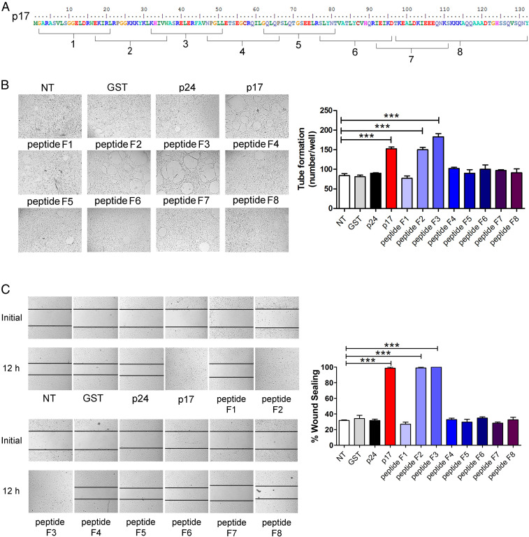
Fig. 1.
Ability of different p17-derived peptides to induce angiogenesis and migration on HUVECs. (A) aa. sequence of eight p17-derived peptides. (B) HUVECs were cultured under stressed condition (EBM containing 0.5% fetal bovine serum [FBS]) for 16 h at 37 °C and then stimulated for 8 h at 37 °C with 10 ng/mL GST, p24, p17, or each p17-derived peptide (F1, F2, F3, F4, F5, F6, F7, F8). NT, not treated. (C) HUVECs were cultured under stressed condition for 16 h at 37 °C, and then the confluent cell monolayers were scratched and stimulated for 12 h at 37 °C with medium alone (NT) or with medium containing 10 ng/mL GST, p24, p17, or each p17-derived peptide (F1, F2, F3, F4, F5, F6, F7, F8). Images are representative of three independent experiments with similar results. (Original magnification, 10×.) Data are the mean ± SD of one representative experiment, of three with similar results, performed in triplicate. Statistical analysis was performed by one-way ANOVA, and the Bonferroni post hoc test was used to compare data (***P < 0.001).

