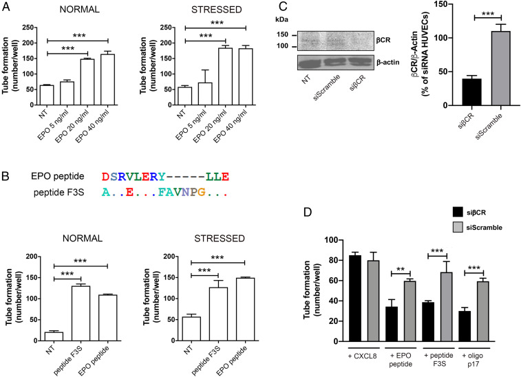
Fig. 2.
EPO peptide and peptide F3-induced angiogenesis is mediated by βCR. (A) HUVECs were cultured under normal (EGM containing 10% FBS) or stressed conditions (EBM containing 0.5% FBS) for 16 h at 37 °C and then stimulated for 8 h at 37 °C with 5, 20, or 40 ng/mL EPO in complete medium. NT, not treated. (B, Upper) Peptide F3 has been modeled on the region of EPO showing the maximum rate of mimicry. (B, Lower) HUVECs were cultured and stimulated as above. NT, not treated. Values reported for tube formation are the mean ± SD of one representative experiment, of three with similar results, performed in triplicate. Statistical analysis was performed by one-way ANOVA, and the Bonferroni post hoc test was used to compare data (***P < 0.001). (C) Western blotting analysis (Left) performed 72 h after nucleofection of HUVECs with βCR siRNA (siβCR) and control siRNA (siScramble). The densitometric data (Right) are corrected by β-actin levels and expressed as percentage of siβCR (means ± SD, n = 4). Statistical analysis was performed by t test, ***P < 0.001 (siβCR vs. siScramble). (D) Seventy-two hours after nucleofection with siβCR or siScramble, HUVECs were stimulated for 8 h at 37 °C with 10 ng/mL CXCL8, EPO peptide, peptide F3S, or oligomeric p17. Values reported for tube formation are the mean ± SD of one representative experiment, of three with similar results, performed in triplicate. Statistical analysis was performed by one-way ANOVA, and the Bonferroni post hoc test was used to compare data (**P < 0.01, ***P < 0.001).

