Figure 1.
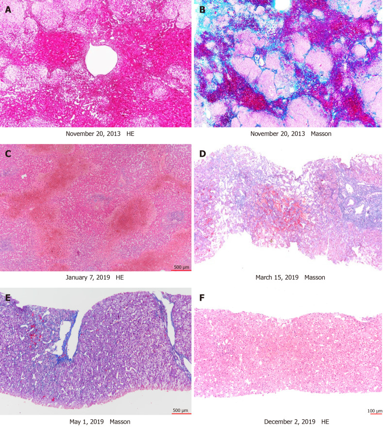
Hepatic venography. A and B: Native liver showed marked sinusoidal dilatation and congestion in centrilobular regions and extensive bridging fibrosis and necrosis linking central to central areas; C: Explanted first liver graft characterized by massive perivenular congestion and hemorrhage with marked sinusoidal dilatation. Portal tract was not remarkable; D: Two months after liver retransplantation, liver biopsy was performed to clarify the diagnosis. The second liver graft liver pathology showed sinusoidal dilatation and congestion; E: In addition to warfarin, tacrolimus was switched to cyclosporine A. Two months after treatment, perivenular congestion and sinusoidal dilation were alleviated and were only observed in the focal perivenular area; F: Nine months later, there was no perivenular congestion and only mild sinusoidal dilatation.

