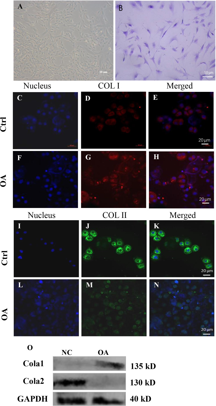
Figure 6. Cell biology detection.
(A) Morphological observation of chondrocytes. (B) Microscopic observation of toluidine blue staining of chondrocytes. (C)–(N) are the expression of COL I and COL II by immunofluorescence staining in human chondrocytes. (O) is the expression of COL I and COL II by fluorescence western blot in human chondrocytes.

