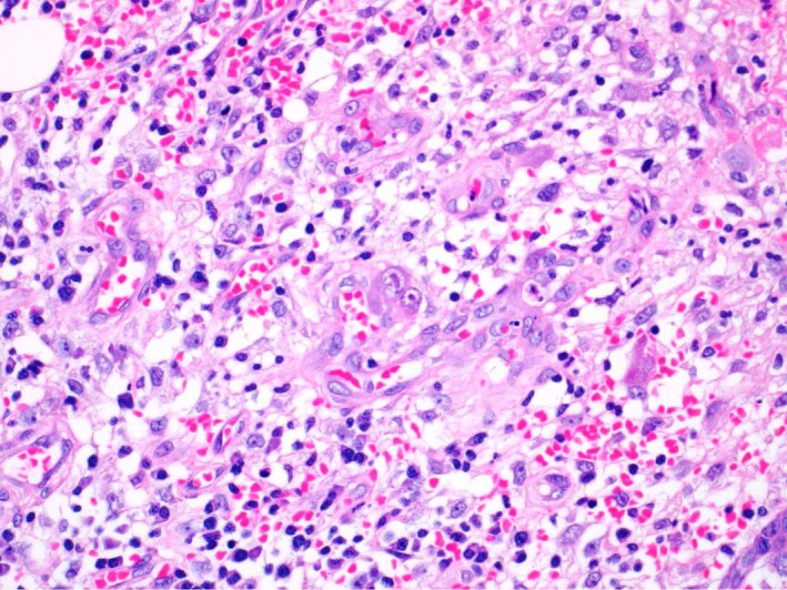
Figure 3.

High power magnification photograph at the perforation site showing mixed acute and chronic inflammation in the lamina properia. In the middle of the photograph, there is enlarged cell with eosinophilic intranuclear and intracytoplasmic inclusions in the lamina properia in the ulcer bed, indicative of cytomegalovirus inclusions
