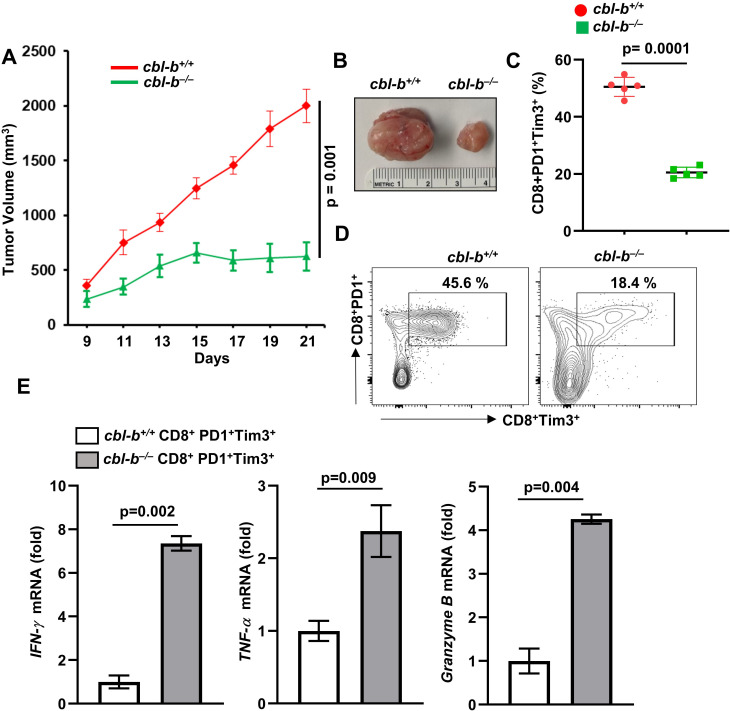
Figure 3.
Reduced MC38 tumor growth in cbl-b–/– mice. cbl-b+/+ and cbl-b–/– mice (n=5) were subcutaneously injected with MC38-CEA cells. (A) Tumor volume. (B) Representative images of tumors. (C) Percentage of CD8+PD1+Tim3+ TILs. (D) Representative image showing percentage of CD8+PD1+Tim3+ TILs. (E) CD8+PD1+Tim3+ TILs were sorted and expression of IFN-γ, TNF-α and granzyme B was analyzed by real-time PCR. The data are representative of three independent experiments. Statistics are mean±SD, calculated by Student’s t-test (two-tailed). CEA, carcinoembryonic antigen; IFN-γ, intergeron gamma; TIL, tumor-infiltrating lymphocyte; TNF-α, tumor necrosis factor alpha.

