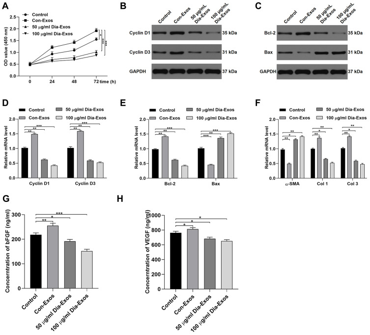
Figure 2.
Dia-Exos suppress the survival and functionality of HSFs in vitro. (A) CCK-8 assays were conducted to measure HSF proliferation following exosome treatment; (B) Western blotting was used to quantify Cyclin D1 and Cyclin D3 protein levels following Dia-Exo treatment; (C) Western blotting was used to quantify Bcl-2 and Bax protein levels following Dia-Exo treatment; following Dia-Exo treatment, qPCR was used to evaluate the expression of (D) Cyclin D1 and Cyclin D3 and (E) Bcl-2 and Bax; (F) The impact of Con-Exo or Dia-Exo treatment on collagen-related marker expression in HSFs was measured to evaluate collagen synthesis; (G–H) ELISAs were used to measure bFGF and VEGFA secretion by HSFs. Data are means ± SD from triplicate experiments. *p < 0.05, **p < 0.01, ***p < 0.001.

