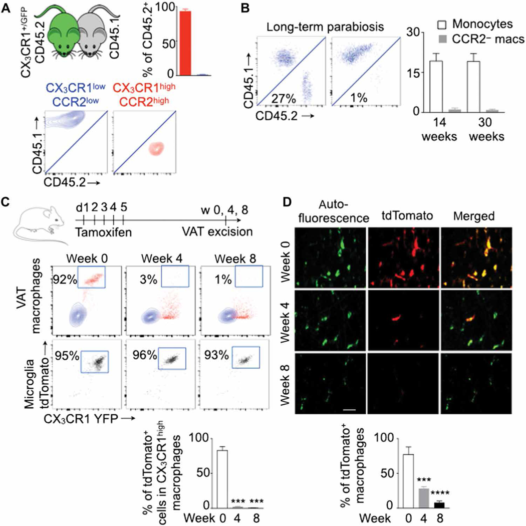
Fig. 1. CX3CR1high CCR2high and CX3CR1low CCR2low macrophages in adipose tissue are monocyte-derived and resident macrophages, respectively.
All experiments were performed in lean transgenic mice without MI. (A) Parabiosis between wild-type (CD45.1) and CX3CR1+/GFP (CD45.2) mice was performed, and visceral adipose tissue (VAT) in CD45.1 mice was analyzed for chimerism 2 months after the parabiosis. The chimerism in VAT macrophage subsets was adjusted to that in blood monocytes. n = 4 pairs per group. (B) Chimerism in blood monocytes and VAT-resident macrophages (macs) was quantified at 14 and 30 weeks after parabiosis. n = 6 pairs per group. (C and D) Genetic fate mapping in CX3CR1CreER/+ ROSAtdTomato/+ mice to test the origin of CX3CR1high CCR2high macrophages. TdTomato+ (CX3CR1high) macrophages were quantified at different time points after tamoxifen injection using flow cytometry (C) and confocal microscopy (D). VAT macrophages are autofluorescent (green). Scale bar, 7 μm. n = 3 to 11 per group. Data are derived from three independent experiments. Means ± SEM. ***P < 0.001 and ****P < 0.0001.

