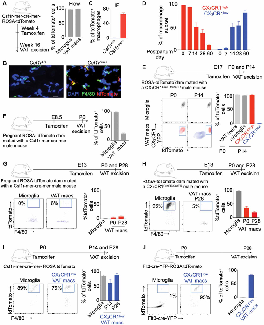
Fig. 2. VAT-resident macrophages are derived from progenitors present at birth.
All experiments were performed in lean transgenic mice without myocardial infarction (MI) at different time points after birth as shown. (A to C) The frequency of tdTomato+ VAT-resident macrophages was quantified using flow cytometry (A) (n = 7 per group) and confocal microscopy (B and C) (n = 3 to 4 per group). Scale bar, 20 μm. (D) Frequency of the VAT macrophages at different time points after birth in CX3CR1+/GFP mice. n = 3 to 4 per group. (E to J) Experimental design and quantification of tdTomato+ VAT macrophages labeled by tamoxifen injection in various lineage-tracing mice. n = 3 to 4 per group in (E); n = 3 per group in (F); n = 5 to 6 per group in (G); n = 5 to 12 per group in (H); n = 4 (P14) and n = 8 (P28) in (I); and n = 3 (microglia) and n = 8 (macrophages) in (J). Tamoxifen was injected in either pregnant dams or offspring, and tdTomato+ VAT-resident macrophages were enumerated by flow cytometry at various time points as shown. Data are from two to three independently performed experiments. IF, immunofluorescence; DAPI, 4′,6-diamidino-2-phenylindole. Means ± SEM.

