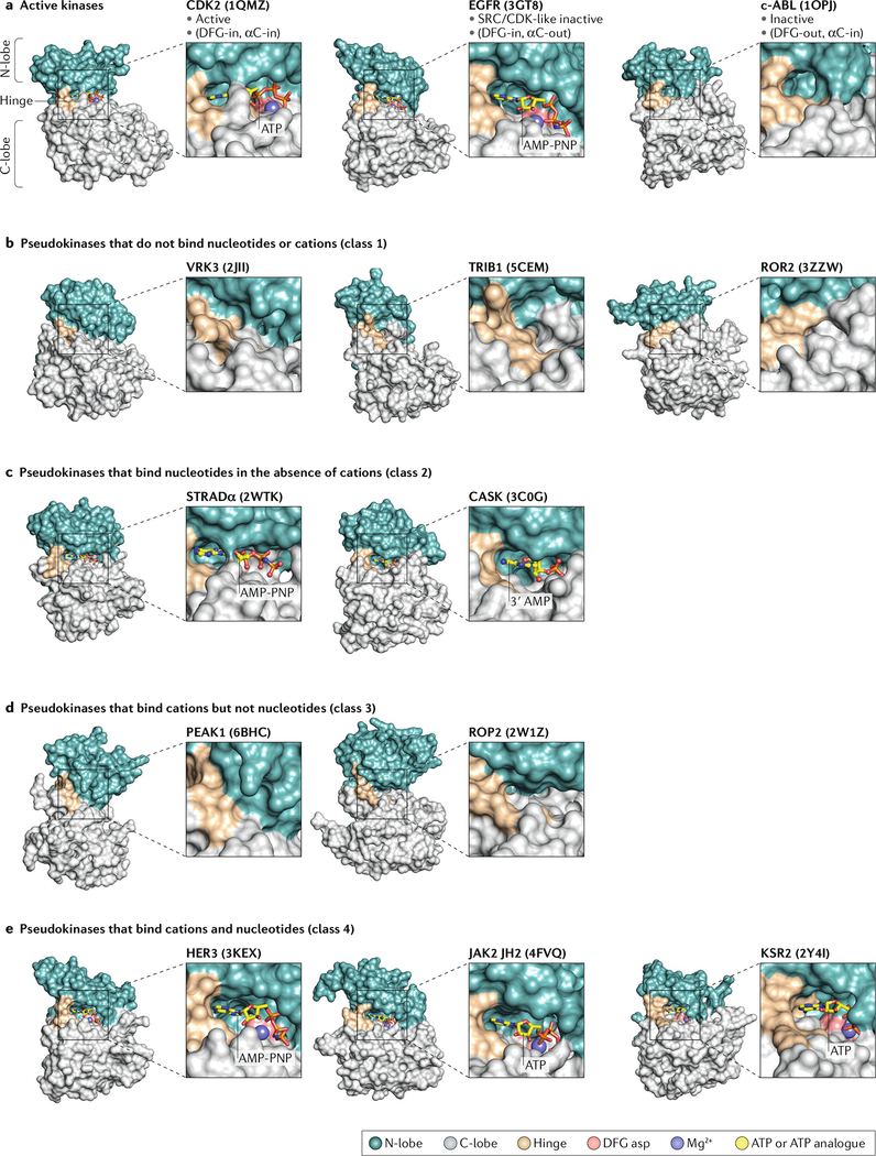
Fig. 4: Accessibility of the putative nucleotide-binding pocket in different classes of pseudokinases.
Surface representations of crystal structures of active kinases in the active DFG-in (CDK2), SRC/CDK-like inactive (EGFR) and inactive DFG-out conformations (c-ABL) (part a), pseudokinases that do not bind nucleotides or cations (part b), pseudokinases that bind nucleotides in the absence of cations (part c), pseudokinases that bind cations but not nucleotides (part d) and pseudokinases that bind nucleotides and cations (part e). For each structure, bound ATP or ATP analogues are shown as sticks, while Mg2+ ions are shown as spheres. Insets show zoomed views of the putative nucleotide-binding pocket for each kinase or pseudokinase. The corresponding Protein Data Bank codes for each crystal structure shown are indicated in parentheses. αC, helix αC; AMP-PNP, nonhydrolysable ATP analogue; C-lobe, carboxyl-terminal lobe; EGFR, epidermal growth factor receptor; HER3, human EGFR; N-lobe, amino-terminal lobe; JAK2, Janus kinase 2; JH2, JAK homology 2; KSR2, kinase suppressor of RAS 2; ROP2, rhoptry protein 2; STRADα, STE20-related adaptor-α.

