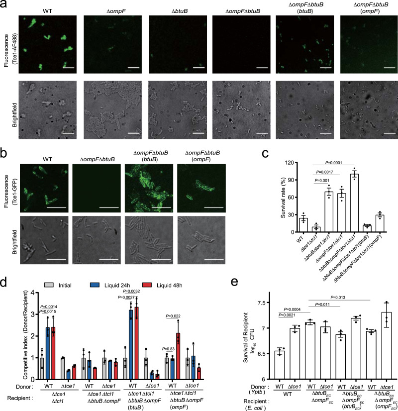
Fig. 4. Tce1 requires BtuB and OmpF for target cell entry.
a, d Fluorescence labeling of the indicated Yptb strains with Tce1-AF488 (a) or GFP-Tce1 (b). Note that the ΔbtuBΔompF mutant shows no labeling, while WT Yptb and complemented strains are labeled in both assays (scale bars, 20 μm). Quantification of a and b was shown in Supplementary Fig. 4c, d. c Toxicity assays of purified Tce1 protein to Yptb strains. The indicated Yptb strains were diluted 40-fold in M9 medium and treated with purified Tce1 (0.1 mg ml−1) for 1 h, and the viability of cells was determined by counting the CFU after treatment. d Intra-species growth competition experiments between the indicated Yptb donor and recipient strains. Donor and recipient strains were mixed 1:1 and grown for 24 or 48 h in liquid medium at 26 °C. Bars represent the mean donor:recipient CFU ratios of three independent experiments (±SD). e Inter-species growth competition experiments between the indicated Yptb donor and E. coli DH5α recipient strains. Donor and recipient strains were mixed 10:1, grown for 12 h in liquid medium at 26 °C. The survival of E. coli cells was quantified by counting CFUs on selective plates. Data are mean ± SD from three biological replicates. P-values from all data were determined using two-sided, unpaired Student’s t-test, and significant differences were considered as P < 0.05.

