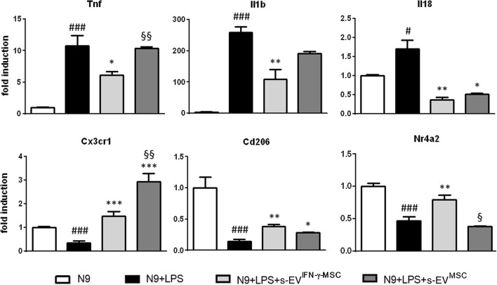
Figure 1.
Exposure to MSC-derived s-EV affects the molecular phenotype of activated microglia. Enhanced anti-inflammatory effect of s-EVIFN-γ-MSC. RT-PCR quantification of genes associated with a pro- (Tnf, Il1b and Il18) and anti-inflammatory/neuroprotective (Cx3cr1, Cd206 and Nr4a2) phenotype in LPS-activated N9 cells. #P < 0.05 (Il18), ###P < 0.001 (Tnf; Il1b; Cx3cr1; Cd206; Nr4a2): untreated (N9) vs LPS-activated N9 cells (N9 + LPS); *P < 0.05 (Tnf; Il18; Cd206), **P < 0.01 (Il1b; Il18; Cd206), ***P < 0.001 (Cxcr3): N9 + LPS vs N9 + LPS exposed for 24 h to s-EVIFN-γ-MSC (N9 + LPS + s-EVIFN-γ-MSC) or s-EVMSC (N9 + LPS + s-EVMSC); §P < 0.05 (Nr4a2), §§P < 0.05 (Cx3cr1): N9 + LPS + s-EVIFN-γ-MSC vs N9 + LPS + s-EVMSC. Data are presented as mean ± SEM of 3 independent experiments conducted in triplicates.

