Figure 6.
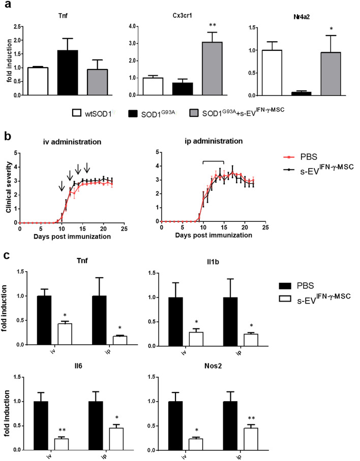
Exposure to s-EVIFN-γ-MSC downregulates neuroinflammation in neurodegenerative disease models in vitro and in vivo. (a) RT-PCR quantification of Tnf and genes associated with an anti-inflammatory/neuroprotective phenotype (Cx3cr1, Nr4a2) in SOD1G93A primary microglia, after a 24-h exposure to s-EVIFN-γ-MSC. Data are presented as mean ± SEM of 3 independent experiments conducted in triplicates. * P < 0.05 (Nr4a2), ** P < 0.01 (Cx3cr1): SOD1G93A vs SOD1G93A + s-EVIFN-γ-MSC. (b) C57BL/6 J mice induced for EAE were treated from the day of disease onset with s-EV administered intravenously or intraperitoneally (n = 8 per group). Data are presented as the mean ± SEM daily clinical score. Arrows represent the times of iv administration (left panel) and the line above the curve represents the daily ip administration (right panel). (c) RT-PCR analysis of pro-inflammatory markers (Tnf, Il1b, Il6 and Nos2) expression was assessed in spinal cords isolated at 20 dpi from EAE mice treated with s-EVIFN-γ-MSC or vehicle (at least n = 4 per group). Results are shown as mean ± SEM of 3 independent EAE experiments with n = at least 3 spinal cord tested per group. *P < 0.05, **P < 0.01.

