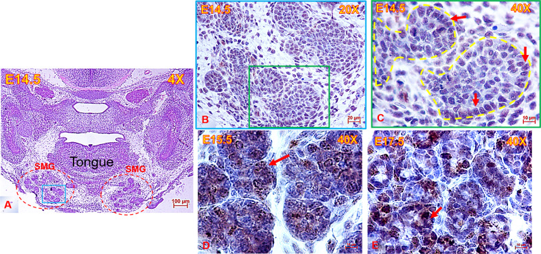
Fig. 1. Anatomic location & histomorphology of the embryonic submandibular glands.
A Low magnification image of E14.5 mouse head (frontal view) stained with H&E and showing the anatomical position of the submandibular glands (SMG) (red dotted outline) beneath the tongue, scale bar = 100 µm. Area outlined with blue square in (A) is stained with AURKB in (B), scale bar = 20 µm. C–E High magnification images showing abundant immunolabelling of nuclei with AURKB (brown) in the mouse SMGs throughout development. AURKB was mainly expressed in the bud nuclei, in addition to sporadic cytoplasmic immunostaining, scale bar = 10 µm.

