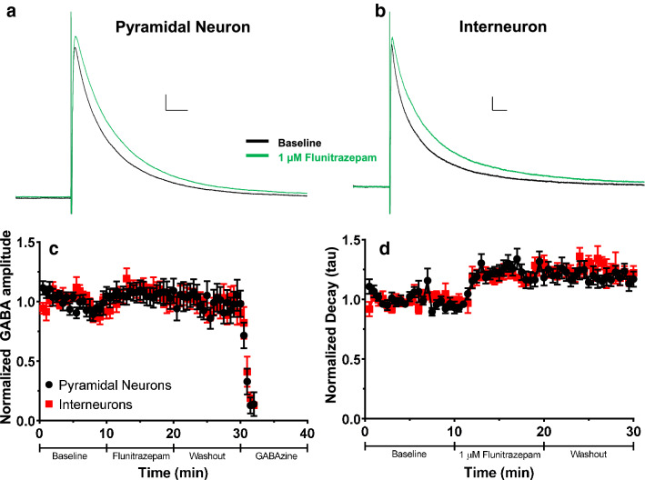
Figure 3.
Effect of acute flunitrazepam application on evoked GABAA receptor-mediated postsynaptic current (GABAA-ePSC) amplitude and decay in layer V pyramidal neurons and INs. (a) Average ePSC traces from layer V pyramidal neurons during the baseline (black trace) and acute 1 µM flunitrazepam application (green trace) phases. Scale bars 40 ms, 50 pA. (b) Average GABAA-ePSC traces from layer V INs during the baseline and acute 1 µM flunitrazepam application phases. Scale bars 40 ms, 20 pA. (c) Normalized GABAA-ePSC amplitudes for both pyramidal neurons (black circles) and INs (red squares) in layer V during the baseline, 1 µM flunitrazepam, washout, and gabazine application phases. (d) Normalized decay constants (tau) for both pyramidal neurons and INs during the baseline, 1 µM flunitrazepam, and washout phases. Data are presented as mean ± SEM.

