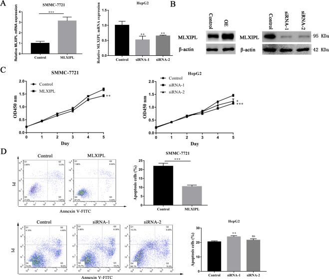
Fig. 6. MLXIPL promotes HCC proliferation and inhibits apoptosis in vitro.
A, B The transfection effect of overexpressed MLXIPL plasmids or MLXIPL siRNAs was measured by quantitative real-time PCR and western blotting. **P < 0.01; ***P < 0.001. C The proliferation ability in indicated cells was detected by the CCK8 assay after MLXIPL overexpression and knockdown separately in HCC cells. *P < 0.05; **P < 0.01; ***P < 0.001. D Apoptosis analysis in indicated cells was detected after MLXIPL overexpression and knockdown separately in HCC cells. Representative data are featured, presenting the population of living cells (Annexin V‑FITC−/PI−) in the left lower quadrant, early apoptotic cells (Annexin V‑FITC+/PI−) in right lower quadrant, late apoptotic cells (Annexin V‑FITC+/PI+) in the right upper quadrant and necrotic cells (Annexin V‑FITC−/PI+) in the left upper quadrant. ns P > 0.05; **P < 0.01; ***P < 0.001.

