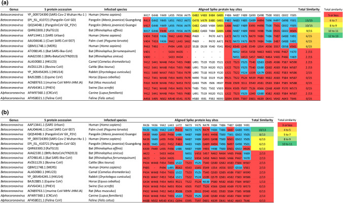
Figure 3.
Amino acid comparisons at key S protein sites among 17 mammalian-specific coronaviruses. (a) Displays the conservation of key ACE2-binding sites on SARS-CoV-2 S protein by 16 mammalian-specific S proteins. (b) Displays the conservation of key ACE2-binding sites on SARS-CoV S protein by 16 mammalian-specific S proteins. Highlighted blue and red are matching and mis-matching amino acid residues, respectively, between mammalian coronaviruses and the two human SARS coronaviruses at key S protein sites. Highlighted yellow is the 482–485 GVEG motif found in SARS-CoV-2. Total similarity designates the total number of matching amino acid residues with respect to SARS-CoV-2 or SARS-CoV, and scores in green highlights high similarity, in light green highlights medium–high similarity, in yellow highlights medium similarity, in orange highlights medium–low similarity, and in red highlights low similarity.

