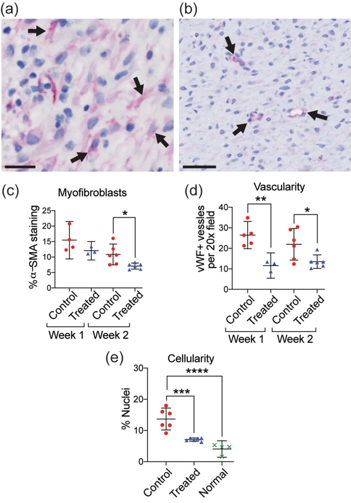
Figure 4.

Reduction in myofibroblasts, vascularity, and dermal cellularity in MSTC-treated wounds. Representative images of immunohistochemical staining for α-smooth muscle actin (a, myofibroblasts highlighted by arrows, scale bar = 25 μm) and von Willebrand factor (b, vascular structures highlighted by arrows, scale bar = 50 μm). MSTC-treated wounds had reduced densities of α-SMA + myofibroblasts after 2 weeks (c), as well as vWF + blood vessels at both time points (d). Overall cellularity in the dermis was significantly elevated in control wounds at 2 weeks, compared to both MSTC-treated wounds and unwounded normal skin (e). *p 0.05, **p 0.005, ***p 0.0005, ****p 0.0001.
