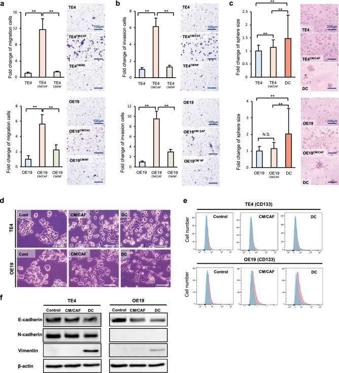
Figure 2.
Tumor cells stimulated with CAFs acquired malignant characteristics. The migration (a) and invasion (b) of esophageal cancer cells cultured with conditioned medium from CAFs were observed and quantified. CAFs enhanced the migration and invasion of cancer cells (scale bar, 100 μm). (c) The results of a spheroid formation assay are shown. Spheroid formation was enhanced by CAFs. The spheroid sizes in the direct-contact (DC) groups were significantly larger than those in the control groups (scale bar, 200 μm). (d) The morphological changes of cancer cells stimulated with CAFs are shown. Compared with untreated control cells, the stimulated tumor cells had a larger population with fewer cell junctions and elongated pseudopodia. These changes were observed more clearly with direct contact between the cancer cells and CAFs (scale bar, 200 μm). (e) The population of CD133-positive cells was analyzed by flow cytometry. A significant difference was not observed in TE4 cells. However, OE19 cells stimulated with CAFs contained more CD133-positive cancer stem-like cells than unstimulated OE19 cells. (f) WB demonstrated that E-cadherin expression was decreased and vimentin expression was increased in CAF-stimulated tumor cells. These changes were observed to be strong in cultures with direct contact between the cancer cells and CAFs. Data are shown as the mean ± SD of three or more independent experiments. Statistical analyses were performed using Dunnett test. **P < 0.01. N.S. indicates no significant difference.

