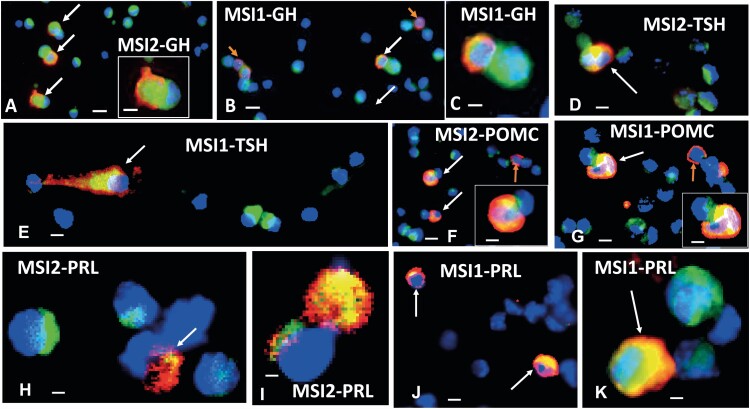
Figure 4.
Musashi expression in dispersed anterior pituitary (AP) cell lineages. Immunolabeling for growth hormone (GH), thyroid stimulating hormone-β (TSH-β), proopiomelanocortin (POMC), or prolactin (Prl; red fluorescence, Dylight 594 or Cy3) and MSI2 or MSI1 (green fluorescence Dylight or Alexa Fluor 488). Blue is DAPI (4′,6-diamidino-2-phenylindole) fluorescence, indicating nuclei. A number of cells are labeled only for MSI2 or MSI1 (green patches near blue nuclei). White arrows show dual-labeled cells. A shows multiple GH cells with MSI2, and one of them is shown in a higher magnification inset. GH is cytoplasmic and MSI is more centrally located, nearer the blue nucleus. B shows some GH cells without MSI1 (orange arrows). C is a higher magnification of 2 cells shown in B, one showing MSI1 and GH (red and green) and the other showing only MSI1. D shows 2 cells immunolabeled for TSH-β and MSI2 next to several cells labeled only for MSI2. E shows a cell dual-labeled for MSI1 and TSH-β, with MSI1 more centrally located. F shows a field with 3 POMC cells, 2 of which are labeled for POMC and MSI2. A third cell (upper right) contains only Pomc (orange arrow). G shows 3 POMC cells, 2 of which are strongly labeled for MSI1. One of the POMC cells is shown in higher magnification in the inset in F and G. H and I show Prl cells dual-labeled for Prl and MSI2 and illustrates the variety in the labeling patterns. J and K illustrate Prl cells dual-labeled for Prl and MSI1. Bar = 10 μm (A, B, D, F, G, and J); 20 μm (insets and C, E, H, I, and K).

