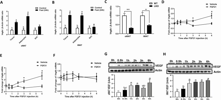
Figure 2.
FGF21 promoted expression and accumulation of VEGF in WAT. The relative mRNA abundance of Vegfa in tissue, adipocytes and stromal vascular fraction isolated from eWAT (A) and iWAT (B) at fed or 24 h of fasting. Twelve-week-old male WT (FGF21fl/fl) and FGF21 LKO mice were fasted for 24 h; the Vegfa mRNA expression in eWAT and iWAT (C). Quantitative reverse transcription PCR analysis for Vegfa mRNA expression in eWAT (D), iWAT (E), and BAT (F) at the indicated time points after tail vein injection of rmFGF21 (1 mg/kg). The protein levels of VEGF at various time points after mice receiving a delivery of rmFGF21 with tail vein injection in eWAT (G) and iWAT (H). n = 6/group. Statistical significance was evaluated by unpaired Student’s t test. *P < 0.05, **P < 0.01, versus control; labeled means without a common letter differ, P < 0.05. Abbreviation: SCV, stromal vascular fraction.

