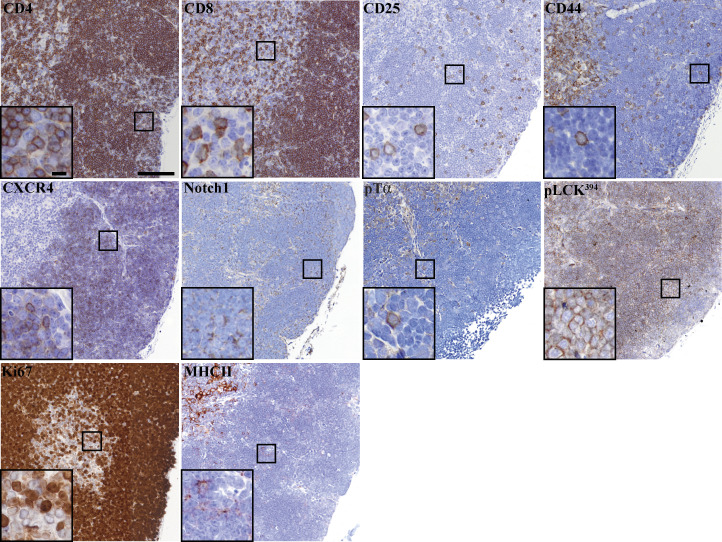
Figure S5.
Chromogenic detection of markers used in Opal multiplex. Thymus lobe slices of 4 µm thickness were mounted on a glass slide, and chromogenic detection (brown) of the multiplex thymus panel markers was performed, followed by counterstaining with hematoxylin to visualize the nucleus (blue). Images were acquired using an Olympus V120 slide scanner, and representative images are shown. Scale bar, 100 µm (10 µm in zoomed images).

