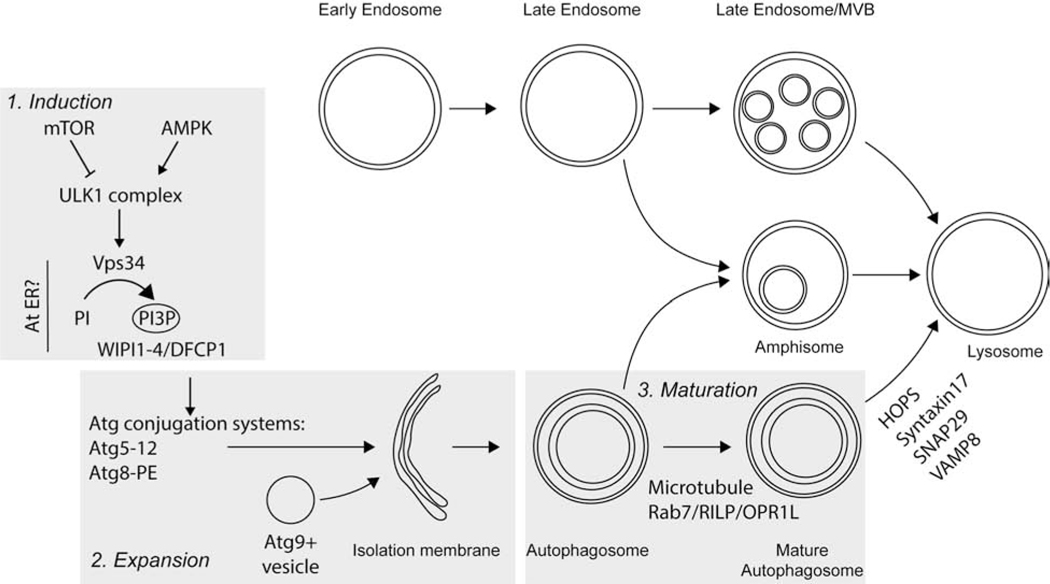
Figure 1. Schematic representation of the mechanisms of autophagy.
Autophagy induction is driven by mTOR and AMPK. These metabolic kinases stimulate PI3P synthesis which recruits the PI3P binding proteins DFCP1 and WIPI1-4 to a membrane source. The activity of the Atg conjugations systems and Atg9+ vesicles expand the autophagic membrane. Once the membrane closes, the autophagosome matures, traffics to the perinuclear area and fuses with lysosomes. Late endosomes can fuse with closed autophagosomes to form amphisomes. Cargo is not depicted for simplicity.

