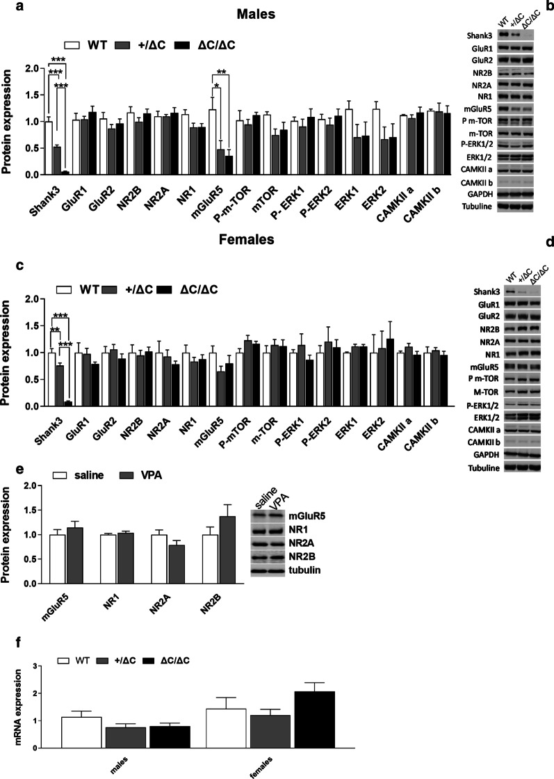
Fig. 7.
Brain levels in PSD proteins and mRNA in ASD mouse models. a–c Expression profile of Shank3 and associated postsynaptic scaffolding proteins in males and females Shank3 mice within the cerebellum by immunoblot analysis. b–d Representative figures of the western blot. Note that only mGluR5 was significantly decreased in Shank3+/ΔC and ΔC/ΔC males compared to wild type. No decreases were present in Shank3 ΔC/ΔC and Shank3+/ΔC females. Experiments for western blots were repeated at least three times and statiscal analysis were performed with a one-way ANOVA followed by Tukey’smultiple analysis (*p < 0.05, **p < 0.01, ***p < 0.001) WT males n = 4; Shank3+/ΔC males n = 8; Shank3 ΔC/ΔC males, n = 8; WT females n = 5; Shank3+/ΔC females n = 8; Shank3 ΔC/ΔC females, n = 9. e No difference in the protein levels of several glutamate receptors was found in the male cerebellum of VPA ASD mouse model whatever the prenatal treatment (Saline, n = 5; VPA, n = 5, Student’s t test, p > 0.05). f No difference in the mGluR5 mRNA levels was found in the cerebellum whatever the genotype or sex (wild type males (n = 7), Shank3+/ΔC males (n = 8), Shank3 ΔC/ΔC males (n = 8), wild type females (n = 7), Shank3+/ΔC females (n = 8), Shank3 ΔC/ΔC females (n = 8), One-way ANOVA, p > 0.05). All data are expressed as means ± SEM

