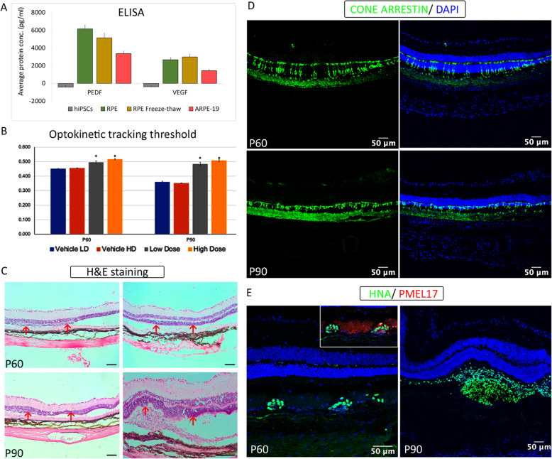
Fig. 6.
In vivo efficacy studies of RPE cells after transplantation into animal models. a ELISA-based quantification of polarization proteins secreted in in vitro culture supernatants—PEDF and VEGF in day 70 RPE running cells and freeze-thawed cells; hiPSCs and ARPE-19 cell line was used as negative and positive controls, respectively. b–e Functional and molecular outcome of RPE transplantation in RCS rats. b Optokinetic tracking thresholds measured from all animals at P60 and P90. Asterisks indicate statistical significance between cell-treated and BSS+-injected controls. c Histological evaluation using H&E staining data at P60 and P90. d Cone arrestin staining in the retina sections at P60 and P90. e Immunofluorescence images show the presence of transplanted cells detected using a human nuclear marker (HNA) co-stained with PMEL17 indicating graft survival and engraftment. Scale bars represent 50 μm

