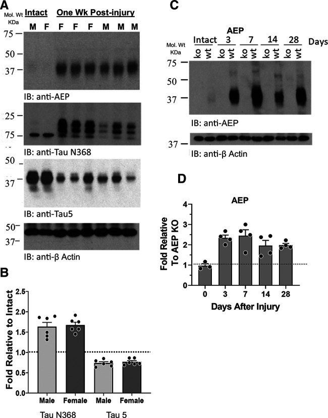
Figure 1.
A, Immunoblots of extracts of sciatic nerves from intact mice and from nerves cut and repaired one week earlier. IR to AEP (top) is noted by the band at 37 kDa, representing enzymatically active AEP. IR to the AEP-cleaved fragment of Tau (Tau N368) and to full-length Tau (Tau 5) are shown below. B, Quantitative analysis of differences in expression of Tau N368 and Tau 5 between intact nerves and cut and repaired nerves, relative to β actin controls, are shown for a group of male and female mice as mean fold (±SEM) intensities. The horizontal dashed line at unity marks the amount of IR found in intact mice. C, Changes in AEP IR are shown at different times after sciatic nerve transection and repair in WT and AEP KO mice. D, Quantitative analysis of AEP expression in sciatic nerves at different times after nerve transection and repair. Mean (±SEM) fold expression of AEP IR, relative to that found in AEP KO mice at the same postinjury times are shown. The horizontal dashed line at unity marks the amount of IR found in AEP KO mice.

