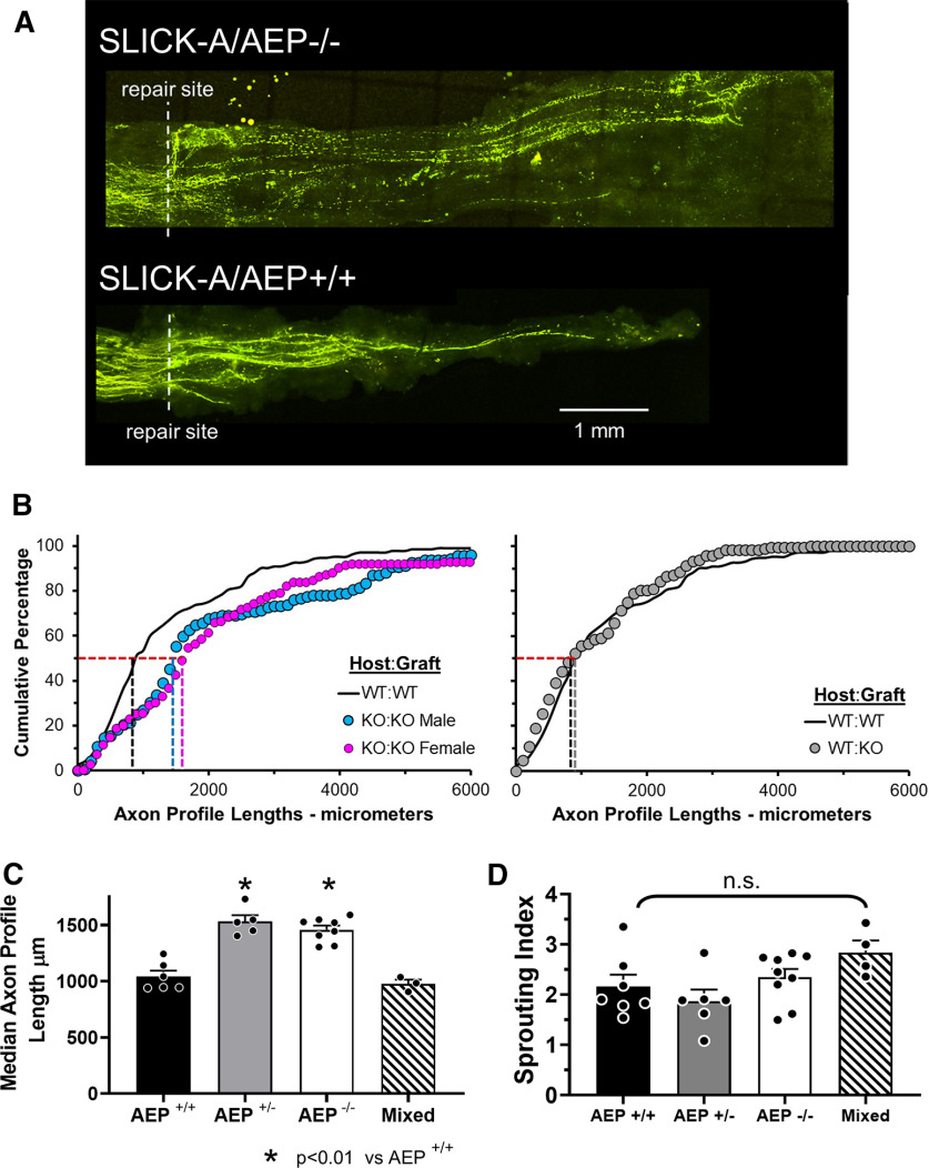
Figure 4.
Elongation of regenerating axons is enhanced in AEP KO mice. A, Examples of confocal images of sciatic nerves in SLICK-A mice that had been cut and repaired two weeks earlier with a segment of nerve harvested from a littermate that did not express YFP. Note more longer axons are found in the SLICK-A/AEP−/− mouse (top). B, Distributions of YFP+ regenerating axon profile lengths in male and female SLICK-A/AEP−/− mice (KO:KO) and SLICK-A/AEP+/+ (WT:WT) mice measured two weeks after sciatic nerve transection and repair are shown as cumulative frequency histograms (left). Data from nerves from WT (SLICK-A/AEP+/+) mice repaired with grafts from AEP−/− mice (WT:KO) in comparison to those from WT:WT nerves are shown in the graph on the right. Each set of symbols represents mean values in each bin (N = 5 except for WT:KO, where N = 4). Vertical dashed lines mark the lengths at the 50th percentile, the median for each distribution. C, Average (±SEM) median lengths of regenerating axon profiles two weeks after sciatic nerve transection and repair in mice in which neither, both, or one copy of the gene for AEP had been knocked out, and when WT nerves were repaired with grafts from KO mice (mixed). D, Mean (±SEM) sprouting index in mice of the three genotypes studied and mixed repair nerves. No significant differences were observed.

