FIGURE 3.

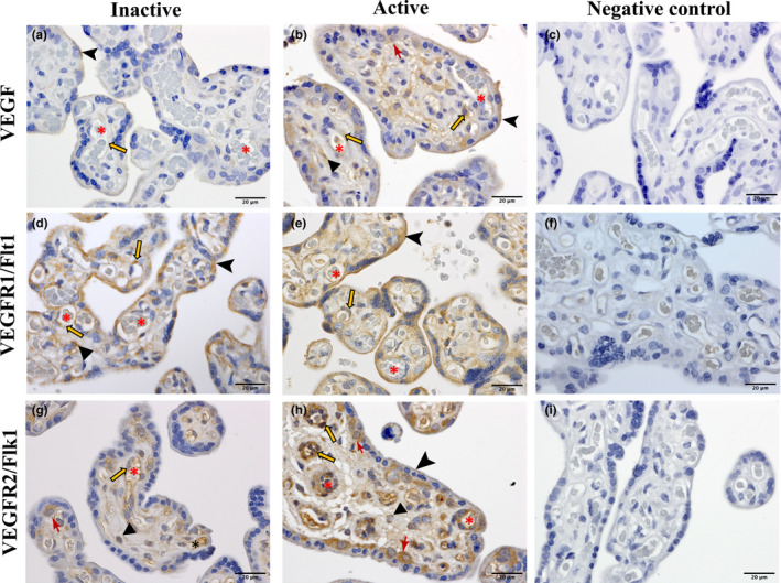
Immunolocalization VEGF (a, b), VEGFR1 (Flt1) (d, e), and VEGFR2 (Flk1) (g, h) in term placenta from physically active and inactive women. c, f, i represent negative controls, performed in absence of primary antibodies (diluent only). red asterisk, lumen of the blood vessels; orange arrows, endothelial cells; triangles, stromal cells; arrow heads, syncytiotrophoblast border, red drafting point arrows, cytotrophoblast cells. All sections were counterstained using hematoxylin. Scale bars, 20 µm.
