Fig. 1.
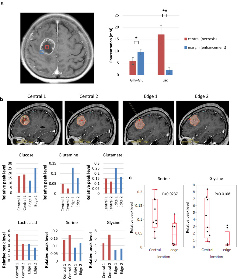
Serine and glycine levels are elevated in the core of tumor with nutrient-poor microenvironment in GBM patients. See also Additional File 2: Supplemental Fig. 1. a Representative MR image for MR spectroscopy (MRS) in a 68-year-old patient with GBM. Volumes of interest (VOIs) of MRS study were placed on the tumor bulk (red square) and the tumor edge (blue square). The relative level of glutamine and glutamate (Gln and Glu), and lactate (Lac) was calculated with respect to creatine and phosphocreatine (Cr and PCr) in 7 GBM patients (statistically significant with *p < 0.05, **p < 0.01). b GC–MS analysis in tumor samples obtained from a 60-year-old patient with GBM. Each sample was targeted in representative MR images using BrainLab navigation system. c Ex vivo serine and glycine levels in four GBM patients (central; 9 samples, edge; 8 samples)

