Fig. 3.
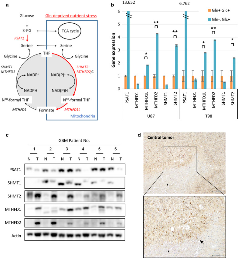
MTHFD2 expressions are elevated in the glutamine-deprived cells and tumor core of GBM patients. See also Additional File 2: Supplemental Fig. 3. a A schematic showing the enzymes involved in one-carbon metabolism that were targeted in this study. PSAT1; phosphoserine aminotransferase 1, SHMT1 and 2; serine hydroxymethyl transferase 1 and 2, and MTHFD1 and 2; methylenetetrahydrofolate dehydrogenase 1 and 2. MTHFD1L; monofunctional tetrahydrofolate synthase, mitochondrial b mRNA levels of PSAT1, SHMT1 and 2, MTHFD1 and 2, and MTHFD1L in U87 and T98 GBM cells which were grown with or without glutamine for 48 h. Data represent the mean ± SEM of three independent experiments (statistically significant with *p < 0.05, **p < 0.01). c Immunoblot analysis of PSAT1, SHMT1 and 2, MTHFD1 and 2 staining in central tumors (T) and normal brain tissues (N) around tumor edge obtained at tumor resection from 6 patients with GBM. d Representative immunohistochemical images of MTHFD2 in central tumors obtained from a GBM patient. Tissue was counterstained with hematoxylin. Scale bar upper 200 μm, lower 100 μm arrow; pseudopalisading asterisk; necrosis

